 |
310-206-2510
|
Breast Brachytherapy
( High Dose Rate Brachytherapy for Breast Cancer )
Back to Top1. Introduction
Click here for a PDF of the CET Breast Brochure.Click here to find out more about Mammosite. Mammosite Patient Stories.
Other helpful Breast Cancer Links.
| Increasingly, women are deciding to have treatment of breast cancer with a safe and effective form of radiation therapy known as "breast brachytherapy". This method of therapy, which delivers radiation directly into a tumor site from the inside out, is a way to save most of the normal breast tissue, preserve the cosmetic appearance of the breast, and avoid the physical and emotional trauma of extensive breast removal surgery. In the past, radiation has been administered to a patient’s “entire” breast (whole breast radiation) via external beam radiation therapy (EBRT). EBRT treatment is delivered to the tumor site via a radioactive beam from outside the patient’s body on a daily basis |
 |
While both external beam and brachytherapy can be utilized to deliver partial breast radiation, brachytherapy is the quickest, most direct, and conformal way to deliver the radiation to the target.
High dose rate (HDR) brachytherapy for breast cancer is usually administered as a complete course given twice a day for a total of 5 days on an outpatient basis. There are two methods of brachytherapy depending upon the size and location of the tumor in relationship to the size and shape of the breast. One is known as Tube and Button and the other is referred to as Balloon Catheter or Mammosite. Both are discussed in more detail below in the Surgery and HDR Procedure section.
Back to TopTable of Contents
- Introduction
- The Advantages of HDR Brachytherapy
- Who's a Candidate for Breast HDR Brachytherapy
- Side Effects
- Surgery and HDR Procedure
- Cosmetic Results of HDR Brachytherapy
- Treatment Planning (Dosimetry)
Back to Top2. The Advantages of HDR Brachytherapy
- Overall treatment time is 1 week versus 6 to 7 weeks for external beam radiation therapy.
- Conserve your breast and yield excellent cosmetic results.
- Breast brachytherapy delivers a precisely targeted dose to the tissues most at risk for recurrence, increasing the likelihood of tumor control.
- Reduces radiation dose to the lungs and opposite breast.
- Avoids potential long term side effects by reducing radiation doses to healthy tissue.
- Breast brachytherapy causes no delays in other treatments such as chemotherapy.
- Placement of the applicator (Tube and Button or Mammosite balloon) is simple and safe.
- Treatment is given on an outpatient basis, so no hospital stay is required.
Back to Top3. Who's a Candidate for Breast HDR Brachytherapy
There are 3 types of breast cancer patients who qualify for HDR brachytherapy:- Patients who have early stage breast cancer.
- Breast Tumor size is 3cm or less (And in some cases 0.5 in or less)
- 0 to 4 lymph nodes positive for disease.
- Patients who have locally advanced breast disease (no metastasis).
- No prior history of radiation treatment for breast cancer.
- Tumors may or may not be fixed to the chest wall.
- Patients who have recurrent breast cancer to the chest wall (these patients may not be a candidate for surgery or choose not to have surgery).
Back to Top4. Side Effects
Some women have experienced minor bruising, redness and discomfort. All of these side effects are common in breast surgery and radiation treatment and usually last 2-4 weeks.Scarring from the tube and button insertion sites or mammosite insertion site decrease and fade over time.
Back to Top5. Surgery and HDR Procedure
The illness must first be diagnosed by biopsy of a breast lump or abnormal area on a mammogram. Then, instead of a mastectectomy (surgical removal of the entire breast), treatment begins with a more limited surgical procedure (lumpectomy) to remove only the abnormal tissue.
Next, selection of the type of local radiation therapy and determination of whether other measures (such as hormones or chemotherapy) are needed. If the tumor is small (3 centimeters or about an inch and a half or less), then only the area where the tumor was removed needs to be treated.
Depending upon the size and location of the tumor in relationship to the size and shape of the breast, your physician will recommend which method of brachytherapy (tube and button or balloon catheter/mammosite) is best suited for you.
- Tube and Button: a series of thin tubes or catheters are placed temporarily through the breast tissue in and around the lumpectomy site. Each catheter is connected to a radiation treatment machine (afterloader) that directs a tiny radioactive source through the tubes and exposes the tissue with radiation according to a predetermined dose plan. This method is technically applicable to virtually any patient who is a candidate for partial breast radiation.
- Balloon Catheter (Mammosite): a small soft balloon attached to a thin cathter tube that's placed inside the lumpectomy cavity (the area where the tissue was removed). The catheter is attached to a radiation treatment machine (afterloader) that directs a tiny radiactive source with millimeter precision into the center the balloon, just as with the tube and button device. The balloon catheter method is most applicable to patients with early breast cancer. The balloon must fit correctly into the region of the lumpectomy and not come too close to the skin's surface.
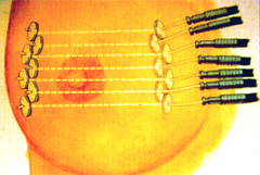 |
Fig. 1-Tube and Button Applicator Insertion Procedure: The tube and button implant can be done at the time of lumpectomy or anytime afterwards. The brachytherapy physician places rows of thin hollow tubes (catheters), to encompass the tumor bed and its margins. The catheters are left in place for five days. Radiographic or CT images of the implant are obtained for treatment planning purposes. Once the radiation dose plan is reviewed and approved by the physician, the treatments can begin. |
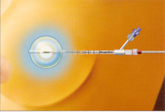 |
Fig. 2-Mammosite Balloon Applicator Insertion Procedure: The surgeon removes the tumor and the uninflated mammosite balloon in inserted into the lumpectomy cavity with a portion of the catheter remaining outside of the breast. The balloon insertion can be done either at the time of the lumpectomy or up to 10 weeks after surgery. Once the balloon is in place it's inflated with a small amount of saline and left in place for five days. Radiographic or CT images of the implant are obtained for treatment planning purposes. Once the radiation dose plan is reviewed and approved by the physician, the treatments can begin. |
Back to Top6. Cosmetic Results of HDR Brachytherapy
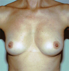 |
Fig. 3-HDR Cosmetic Result: Effects from the catheter placement fade over time and do not cause scarring, dimpling or retraction of the breast tissue. Long term follow up data has shown that accelerated partial breast irradiation is a safe and effective form of breast cancer treatment. Local control rates of breast cancer are equivalent to the control rates found in whole breast external beam radiation treatment (EBRT). In the partial breast irradiation program, surrounding normal breast tissue receive less radiation, and it can be done in 1 week as opposed to 7 weeks.* |
*Results vary by patient
Back to Top7. Treatment Planning (Dosimetry)
 |
||
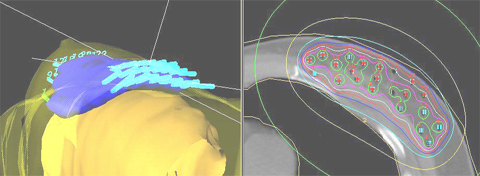
| Fig. 4-3D computer simulation showing a conformal radiation dose cloud: The image on the left is the 3-D representation of the implant catheters (light blue), the 100% radiation dose "cloud" (blue) the underlying lung (yellow), and the skin (transparent yellow). The image on the right is a transverse CT cut showing the levels of radiation dose. Note how the dosimetrist has shaped the treatment volume to avoid the underlying ribs and lung and keeps the skin dose at 50% of the prescription dose to avoid skin reactions. | ||
Home
Treatment Programs
- Prostate Cancer

 Monotherapy Survival Rate
- Breast Cancer
- Vaginal Cancer
- Cervical Cancer
- Endometrial Cancer
- Vulvar cancer
- Head and Neck Cancer
- Lung Cancer
- Esophageal Cancer
- Bile Duct Cancer
- Soft Tissue Sarcoma Cancer
Treatments Statistics
Glossary
Frequently Ask Questions
General Information
Brachytherapy Publications
Cancer Resources & Links
Back to TopGeneral Frequently Asked Questions
1. What is Brachytherapy?
The prefix "brachy" is the Greek word for "short" distance. Brachytherapy is a form of internal radiation treatment where radioactive sources are placed on or into cancer tissues. There are two kinds of brachytherapy. The radiation sources may be inserted either permanently or temporarily. The two most common forms of treatment are low dose rate (LDR) permanent seeds for prostate cancer and high dose rate (HDR) temporary brachytherapy, that can be used for prostate, gynecologic, breast, head and neck, lung, esophageal, bile duct, anorectal, sarcoma, and other cancers.
2. What is high dose rate (HDR) Brachytherapy?
High dose rate (HDR) is a technically advanced form of brachytherapy. A high intensity radiation source is delivered with millimeter precision under computer guidance directly into the tumor killing it from the inside out while avoiding injury to surrounding normal healthy tissue. For a more in depth explanation please visit the understanding HDR Brachytherapy page.
3. How does radiation kill cancer?
Cancer is made of abnormal cells that tend to grow without control. Cancer DNA is more sensitive to radiation than are normal cells, so radiation kills cancer directly or when the cells attempt to multiply while normal tissue in the region is able to repair and recover.
4. What are the advantages of HDR Brachytherapy?
- Short course of treatment compared to other types of radiation treatment (1 week)
- Preservation of organ structure and function
- Fewer side effects
- Excellent coverage of possible microscopic extension of cancer
- Knowledge of radiation dose distribution before treatment is given
- Accuracy and precision of tumor specific radiation dose delivery
- Minimizes areas of radiation overdose (hot spots) or underdose (cold spots)
- Organ motion (target movement) is not a problem for HDR as it is with external beam
| Prostate Specific |
- Effective treatment for cancer recurrence (termed "salvage" therapy)
- No radiation source (seeds) migration into other organs
- No radiation exposure to other persons
| Breast Specific |
- Conserves the breast and yields excellent cosmetic results
- Reduces radiation dose to the heart, lungs, and opposite breast
- Doesn't cause a delay in other treatments such as chemotherapy
For more information on the advantages for specific cancer sites please click on the appropriate link below:
Prostate cancer | Breast Cancer | Gynecologic Cancer | Head & Neck Cancer
Esophageal and Bile Duct Cancer | Lung Cancer | Soft Tissue Sarcoma Cancer
5. How successful is HDR Brachytherapy?
HDR Brachytherapy is effective treatment of local disease in many forms of cancer including prostate, gynecological, breast, head and neck, esophagus, lung, anorectal, bile duct, sarcoma, and other primary cancer or localized metastasis as reported in medical literature. CET's publication on prostate cancer, for example has demonstrated 90% 10-year tumor control. Success rates for other tumors vary according to the type and stage of cancer being treated.
6. How many treatments has CET administered?
As of 12/31/2009, CET has performed 10,267 HDR implants and delivered 21,878 HDR treatments. Please see our treatment statistics for further details.
7. Why is HDR less well known than other forms of cancer treatment?
HDR Brachytherapy is a relatively new form of advance radiation technology. Fewer physicians have been trained to perform HDR procedures compared to seed implants or external beam radiation. Few centers, other than CET have been dedicated to the development of HDR brachytherapy to its full potential. Dr. Demanes has devoted his career to the advancement of brachytherapy and has pioneered the use of HDR and established CET as a center of excellence with specially trained and experienced staff and physicians.
8. Why should I select CET?
- Most experienced HDR center in the country
- First center specializing solely in HDR brachytherapy
- Recognized as HDR experts by colleagues in radiation oncology
- Acknowledged safety record
- Highly trained and experienced physicians and staff
- Long term results published in peer reviewed medical literature
- Quality patient care and follow up
Back to TopAbout Us
-
California Endocurietherapy Cancer Center (CET) is the first brachytherapy only center in the United States.
-
Founded by D Jeffrey Demanes M.D. in 1981.
-
Dedicated solely to High Dose Rate brachytherapy (HDR) since 1991.
-
Most experienced HDR brachytherapy center.
-
A training destination for physicians and residents.
-
HDR treatment protocol development
-
Innovation in high dose rate brachytherapy and equipment design
-
Dedicated to long-term follow-up, outcome studies, and publications in medical journals.
Membership and affiliations
 |
 |
 |
| American Society for Therapeutic Radiology And Oncology Chair - Health Policy and Economics Practice Management Subcommittee, Chair - Regulatory Subcommittee, Member - Health Policy and Economic Committee, Member - Health Policy and Economics Code Development and Valuation Subcommittee, Member - Code Utilization and Application Subcommittee. American Brachytherapy Society Chair - Socioeconomic Committee. American College of Radiation Oncology President - 2005 to 2007 American College of Radiology Fellow - 2007 |
Back to Top
CALIFORNIA ENDOCURIETHERAPY AT UCLA
Division of BrachytherapyDepartment of Radiation Oncology
200 UCLA Medical Plaza, Suite B265
Los Angeles, CA 90095-6951
Tel: 310-206-2510 Fax: 310-794-9795
Hours: Mon-Fri, 8:30AM to 5:00PM Pacific Time
Copyright California Endocurietherapy Medical Corp. All Rights Reserved. www.cetcancercenter.com